Opening Hours: Mon - Fri: 9 am - 9 pm. Saturday by appointment. Sunday Closed.
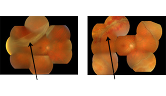
These pair of photos show that Vitrectomy helps patient with Giant Tear and Retinal detachment. This patient has Detachment of Retina with Giant Retinal tear with impairment of vision as shown in the left photos. The right photo shows nicely corrected problem with improvement in vision following Vitrectomy Surgery.
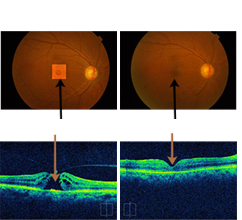
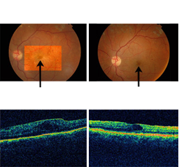
These pair of photos show usefulness of Vitrectomy in Macular hole.This patient has diminished vision in right eye with Macular hole in right eye as seen in fundus photo and confirmed by OCT picture. The right photo shows closed Macular hole as also seen on OCT with improvement in vision following Vitrectomy surgery.
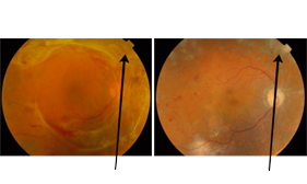
These photos show that Vitreous surgery is the answer in advanced Proliferative Diabetic Retinopathy. This patient has impairment in vision due to scar tissue in retina as a result of Diabetic Retinopathy. The left photo shows postoperative picture after removal of the scar tissue and maintenance of vision.
These photos show that surgical correction of Macular Pucker is a good option. This patient has Macular Pucker that is scar tissue over Macula as seen on OCT and postoperative photo shows improvement in vision after removal of scar tissue.
Please enter your details below to book an appointment.
A sculptor and a Retina specialist have one thing in common that both of them give beautiful vision with their art to a seemingly inconspicuous beginning.
With his training at world renowned Sankara Nethralaya, with ever increasing base of patients of all strata; Dr. Vatsal Parikh decided to move from his earstwhile clinic at Pedder Road to a better and bigger place and this gave the birth to present Drushti Eye & Retina Centre located at Opera House, South Mumbai.
Over the years the centre has earned the reputation of being the best retinal centre for its scientific work with humane touch where even fellow eye surgeons as well as retinal surgeons send their complicated cases for treatment.
Complete eye check up includes detailed Slit lamp examination, detailed Slit Lamp examination, detailed, retinal examination; introcular pressure measurement besides vision and computerised spectacle checking. If required, various specialized investigation
" A stitch in time saves nine " - Diabetic retinopathy is best treated when patient has no complaint but the disease process has already started, in such cases screening and timely treatment is both highly effective and very important. Treatment i
It proves to be a great help in diagnosis and treatment of a variety of retinal conditions by demonstrating various features which are not visible on clinical examination. It also documents the retinal conditions for future progress. Indocyanine Gree
This is the latest & first imaging technique that provides minutest information(upto 10 microns) regarding the retinal layers using laser technology. OCT is most commonly used to diagnose macular disorders like macular hole, macular pucker, diabetic
Laser treatments have been a great help in the prevention and treatment of diabetic patient and various retinal disorders. It is an OPD procedure, recovery is quick and one can resume work immediately after.
Modern vitreo retinal techniques are now available to help treat patients who suffer from advanced vitreo retinal disorders. The technique has brought vision to even those patients that had lost all hopes of being able to see clearly again.
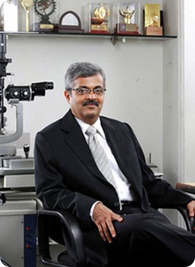
Dr. Vatsal Parikh is a trained vitreoretinal surgeon and eye specialist with 35 years of experience. It is interesting to note that he became retina specialist as his father suffered retinal detachment in 1969 and underwent 10 eye operations in both eyes. This made him experience the plight of retina patients and made his decision to become retina specialist and serve the patients and do research in this field.
M.S., D.O.M.S., F.C.P.S.
Dear Doctor
Thank you very much for the correction and replacement of Cataract Lens in my Right Eye which was spoiled during the first cataract eye... read more.
After many meetings and consulting Dr. Vatsal Parikh operated on me. I couldn't resist myself to put a few words in here for him. Initially... read more.
Paragji Dahyabhai DesaiDear Dr. Parikh
It is no secret that we, that is my mother, husband and my brother, are all quite happy with the cataract operation you... read more.
Dear Dr. Parikh.
We recall our visit to your clinic On a reference from Dr. Mahatmey of Nagpur for retinal examination... read more.